
Apoptosis is the managed or orderly programmed death of a cell.
It is a key process for maintaining cell homeostasis and controlling cell autonomy (Jorgensen et al., 2017).
It was a term introduced by Kerr, Wyllie and Currie to distinguish this type of cell death from necrosis. The term is a combination of two words from the Greek; Apo meaning ‘separation from’ and ptosis meaning dropping or falling down.
The Cell Cycle
The cell cycle is an orderly recurring event which is managed and regulated through a series of genes responsible for both interphase and mitosis. Each cell is regularly dividing. If any cell stays in any phase during the division cycle it is unable to enter the next phase. When cell division is prevented, cells simple do not grow and proliferate.
Encouraging prevention of cell division is a goal for researchers in preventing cancers from growing.
The Nature Of Apoptosis
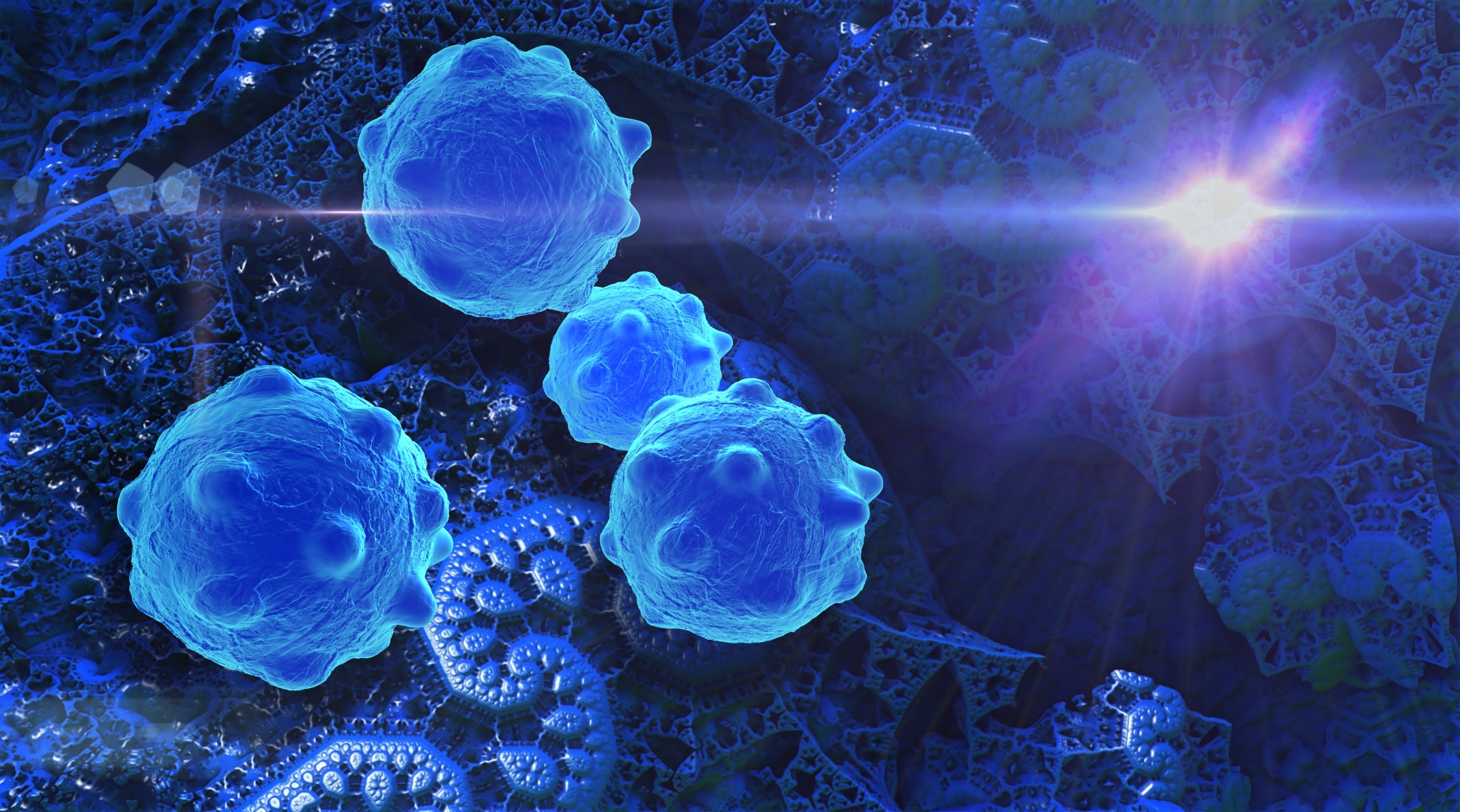
Apoptosis involves the following processes of chromatin condensation, cell shrinkage, the preservation of organelles and cell membranes. There is a rapid engulfment of the cell by neighboring cells to prevent inflammation. Necrosis which is more haphazard involves cell and nuclear swelling. The organelles are disrupted. The cell then ruptures releasing its cellular contents. This is a common case in the inflammatory response.
The origin of programmed cell death (PCD) is ancient. Mitochondria for example have kept their own molecules for triggering cell suicide. These must have been an ancient mechanism which stabilised the organelle as a cell when it first entered the eukaryotic cell. The whole programme of cell death is managed on receipt of a series of apoptotic signals.
The reason for apoptosis is to ensure there is proper development of physiological structures and maintain homeostasis of the whole organism.
Initiation Of Apoptosis
Firstly, a series of events will trigger the process. This can include the following:-
- DNA damage in the cell itself
- Ionizing radiation
- stress and infection by a virus
- cells of the immune system having fulfilled their role and function
- cancerous cells
There are two types of pathway for induction which are either mediated by the extrinsic one of a cell surface death receptor or the intrinsic one which is mitochondrial initiated.
Caspases
A family of proteins which are the main executors of the apoptotic process. They belong to a group of enzymes called the cysteine proteases. They are usually present in all cells as their inactive form (zymogens) and called procaspases. They cleave proteins at aspartic acid residues.
The leading types are caspase 8 and 10 which are initiators in the extrinsic process whilst caspase 9 is an initiator in the intrinsic process. They lead to the production of effector enzymes called caspase 3 and 6 that then cleave key cellular proteins.
The caspase protein structure is a two sub-unit structure with a large 20kD subunit and a smaller subunit of about 10kD.
Other key proteins include those of the Blc-2 family which regulate the activation of the procaspases. They are the inhibitors Bcl-2 and Bcl-Xl. There are also promoters called Bad, Bax and Bak.
The Role Of The Bcl-2 Protein
The Bcl-2 protein is one of the most important regulatory proteins in all the apoptosis family. This protein is located on the outer membrane of mitochondria. It operates in at least three levels of apoptosis (McClintock et al., 2002).
The Extrinsic Mechanism Of Cell Death.
On the surface of all cells are Death Receptors that transmit apoptotic signals which are initiated by particular signaling ligands. The ligands are the following:-
- fas ligand
- Tumor Necrosis Factor (TNF)- alpha
- Tumor necrosis (TNF) -related apoptosis-inducing ligand (TRAI-L)
The signaling ligands activate a caspase cascade within just a few seconds of ligand binding. Induction of apoptosis is extremely quick.
The ligand activation involves production of ceramide which leads to a large number of death receptors clustering together. This helps reinforce and strengthen the apoptotic signal.
In what is called the death domain, there are conformational changes in the intracellular domain of the receptor. The change allows for recruitment of an assortment of apoptotic proteins to the receptor.
The Death Inducing Signalling Complex
The complex is formed from an activating ligand attached to the TNFR1 receptors that cross the plasma membrane and are linked to the Death Domain made up of proteins called TRADD and FADD which are then connected to TRAF-2 and pro-Caspase 8. The protease caspase 8 is activated from its zymogen and apoptosis begins.
Monitoring Apoptosis
The process of apoptosis is measured and monitored using a variety of biochemical techniques. These include:
- The DNA laddering assay
- Phase contrast microscopy
- a determination of ADP/ATP ratios
- The Tunnel assay which is more accurately called the TdT mediated dUTP nick end labelling assay.
In conclusion, apoptosis is an important mechanism for managing cell death. The ramifications are fascinating because it is a point where it is possible to use pharmaceutical intervention. One of the main areas of interest is cancer prevention.
References
, , & (2017). Programmed cell death as a defence against infection. Nature Reviews Immunology, 17(3), 151 (Article).
, , , , , , … (2002). Bcl‐2 Family members and functional electron transport chain regulate oxygen deprivation‐induced cell death. Molecular and Cellular Biology, 22(1), pp. 94–104 (Article).

Leave a Reply