
G Protein-Coupled Receptors (GPCRs), often referred to as seven-transmembrane receptors due to their characteristic structure, constitute the largest and most diverse families of cell surface receptors (Wang et al., 2018). The G proteins they refer to in the term are guanine nucleotide-binding proteins which are used for transmitting signals and function as molecular switches.
Their study has been the subject of a Nobel Lecture by Lefkowitz (2013).
GPCR receptors play a pivotal role in mediating cellular responses to a myriad of extracellular stimuli, including hormones, neurotransmitters, and sensory cues. Their fundamental importance in cellular signaling pathways makes them prominent targets for drug development and therapeutic intervention (Hauser et al., 2017).
This article aims to elucidate the structural organization, signaling mechanisms, and physiological significance of GPCRs, shedding light on their indispensable role in orchestrating cellular responses.
Other Names
The scientific literature sometimes refers to them as as seven-transmembrane domain receptors, 7T receptors, serpentine receptor, and G protein-linked receptors (GPLR) .
Structural Organization of GPCRs
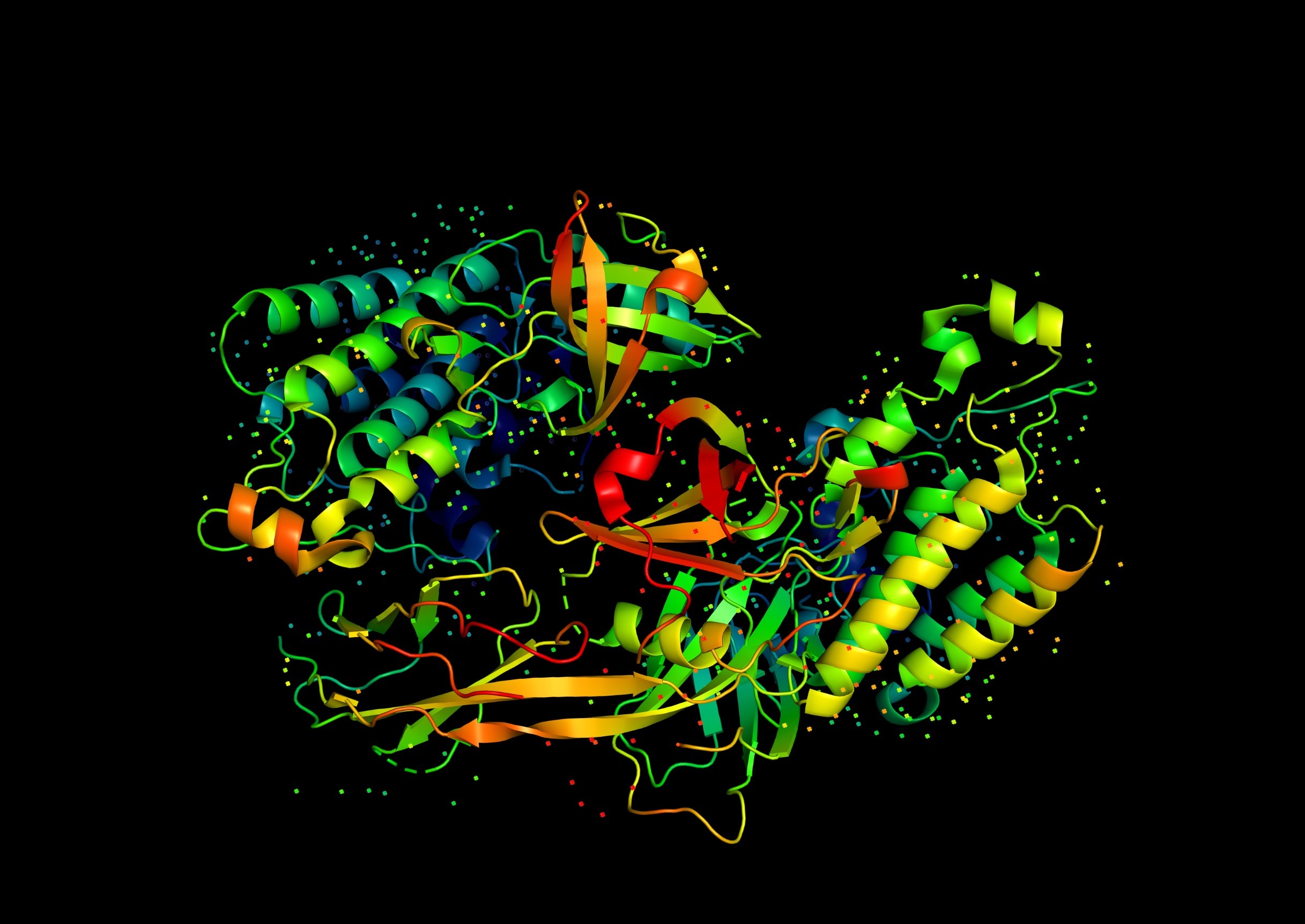
At the core of GPCR structure lies a characteristic seven-transmembrane helical domain, which traverses the lipid bilayer (i.e. the plasma membrane) of the cell membrane. This structural motif forms the basis of the receptor’s ability to transduce extracellular signals into intracellular responses. The structure of a basic GPCR has three distinct parts:
- The extra-cellular region consists of the N terminus and the three extracellular loops (ECL1, ECL2 and ECL3)
- The TM region which are the seven alpha-helices labelled TM1 to TM7.
- The intracellular region which is composed of three intracellular loops (ICL1 to ICL3), an intracellular amphipathic helix (H8) and then the C terminus.
GPCRs exhibit considerable diversity in their primary amino acid sequences, allowing them to recognize and respond to a wide array of ligands. Additionally, the extracellular N-termini and intracellular C-termini, often contain sites for post-translational modifications and interactions with intracellular signaling molecules.
We humans express at least 800 GPCRs and they make up the third largest family of genes.
GPCR Signaling Mechanisms
The signaling mechanisms employed by GPCRs are remarkably intricate and versatile, allowing for fine-tuned regulation of cellular responses. Upon ligand binding to the extracellular domain of the receptor, conformational changes occur within the seven-transmembrane domain, leading to the activation of intracellular signaling cascades.
Central to GPCR signaling is the interaction with heterotrimeric G proteins, which are composed of α, β, and γ subunits. These G protein complexes are made up of 23 alpha, 7 beta and 12 gamma subunits. The beta and gamma subunits can form a stable dimeric complex called the beta-gamma complex.
The alpha subunits form four families labelled Gs, Gi, Gq and G12/13. These are responsible for coupling GPCRs to various distinct effectors. In their inactive state, the α subunit of the G protein is bound to GDP.
We can describe each of the three regions as the extracellular region which modulates access by a ligand. The TM region that forms the structural core binding the ligand and transducing this binding as a message to the intracellular region through conformational change. The intracellular region is the interface with the cytosolic signaling proteins.
What Happens When A Ligand Binds?
Ligand binding induces a conformational change in the GPCR, facilitating the exchange of GDP for GTP on the α subunit of the G protein(s), thereby promoting its dissociation from the βγ subunits. Both the α-GTP and βγ subunits can then modulate the activity of various effector proteins, such as adenylyl cyclase, phospholipase C, and ion channels, leading to the generation of second messengers such as cyclic AMP (cAMP), inositol trisphosphate (IP3), and diacylglycerol (DAG). These second messengers, in turn, initiate downstream signaling events, culminating in cellular responses such as gene transcription, ion channel modulation, and cytoskeletal rearrangements.
GPCR Classification
Classification is based on sequence homology and functional similarity.
Class A or 1..Rhodopsin like
Class B or 2..Secretin receptor family
Class C or 3 ..Metabotropic glutamate and pheromone
Class D or 4 ..Fungal mating pheromone receptors
Class E or 5..cAMP receptors
Class F or 6…Frizzled and/or Smooth.
Another form of classification is based on phylogenetic origin which is the GRAFS system:
- Glutamate
- Rhodopsin
- Adhesion
- Frizzled/taste
- Secretin.
Organization of GPCRs in Cellular Signaling Networks
The spatial and temporal organization of GPCRs within cellular signaling networks is crucial for the integration and coordination of diverse physiological processes. GPCRs exhibit both specificity and promiscuity in their ligand binding properties, allowing them to engage in complex crosstalk with other signaling pathways. Moreover, the regulation of GPCR activity is finely tuned by a plethora of mechanisms, including receptor desensitization, internalization, and recycling, as well as heterologous and homologous receptor regulation. These regulatory mechanisms ensure the precise control of GPCR-mediated signaling in response to changing extracellular conditions and ligand concentrations. Additionally, the existence of GPCR heteromers, wherein two or more distinct GPCRs form functional complexes, further expands the repertoire of signaling outputs and contributes to the diversity of cellular responses elicited by GPCR activation.
Physiological Significance of GPCRs
The physiological significance of GPCRs spans a wide spectrum of biological processes, ranging from neurotransmission and hormone secretion to immune responses and sensory perception. Neurotransmitter receptors, such as the adrenergic, dopaminergic, and serotonin receptors, mediate synaptic transmission and modulate neuronal excitability, thereby regulating mood, cognition, and behavior. Hormonal receptors, including those for adrenaline, insulin, and glucagon, play essential roles in metabolic regulation, energy homeostasis, and stress responses. Moreover, GPCRs involved in immune cell activation and chemotaxis contribute to the inflammatory response and host defense mechanisms. Sensory receptors, such as those for vision, olfaction, and taste, enable organisms to perceive and respond to environmental stimuli, facilitating survival and adaptation to diverse ecological niches.
Signal Molecules
- Amino acids: Glutamate, GABA
- Metal Ions: calcium.
- Peptides & Proteins: insulin, glucagon
- Lipids: prostaglandins, leukotrienes
Targeting GPCRs in Drug Discovery and Therapeutics
The pivotal role of GPCRs in mediating cellular responses and their implication in various pathological conditions make them attractive targets for drug discovery and therapeutic intervention. Approximately one-third of all prescription drugs target GPCRs, highlighting their pharmacological relevance. When Hauser et al., (2017) had written their review there were 475 drugs that acted on 108 unique GPCR targets. That number has increased and there are many passing through clinical trials. Small molecule ligands that modulate GPCR activity by binding to either orthosteric* (see below references) or allosteric sites have been developed to treat a wide range of diseases, including cardiovascular disorders, psychiatric illnesses, and inflammatory conditions.
Moreover, biologics such as monoclonal antibodies and engineered GPCR ligands offer alternative strategies for targeting GPCRs with high specificity and efficacy (Wang et al., 2018; Hauser et al., 2021).
Future Directions and Challenges
Despite the significant advancements in our understanding of GPCR structure, signaling, and pharmacology, several challenges and opportunities lie ahead. These receptors are the targets of a great deal of drug discovery and manipulation. We should see significant progress in our understanding not only of taste but how we manage appetite and metabolism.
Elucidating the structural dynamics of GPCR activation and signaling complexes at atomic resolution remains a formidable task, necessitating the development of innovative experimental and computational approaches. Additionally, unraveling the functional implications of GPCR heteromerization and allosteric modulation poses fundamental questions that warrant further investigation. Furthermore, the emergence of biased signaling, wherein ligands selectively activate specific signaling pathways downstream of GPCRs, presents both challenges and opportunities for drug discovery and precision medicine.
The G Protein-Coupled Receptors (GPCRs) represent a fascinating class of cell surface receptors that govern a myriad of physiological processes through intricate signaling mechanisms. Their structural diversity, signaling versatility, and pharmacological relevance underscore their significance as key regulators of cellular function and attractive targets for drug discovery. Continued research efforts aimed at unraveling the complexities of GPCR signaling and function hold promise for advancing our understanding of cellular physiology and developing novel therapeutic interventions for a wide range of diseases.
References
Hauser, A. S., Attwood, M. M., Rask-Andersen, M., Schiöth, H. B., & Gloriam, D. E. (2017). Trends in GPCR drug discovery: new agents, targets and indications. Nature Reviews Drug discovery, 16(12), pp. 829-842 (Article)
Hauser, A. S., Kooistra, A. J., Munk, C., Heydenreich, F. M., Veprintsev, D. B., Bouvier, M., … & Gloriam, D. E. (2021). GPCR activation mechanisms across classes and macro/microscales. Nature Structural & Molecular Biology, 28(11), pp. 879-888.
Lefkowitz, R. J. (2013). A brief history of G-protein coupled receptors (Nobel Lecture). Angewandte Chemie International Edition, 52(25).
Wang, W., Qiao, Y., & Li, Z. (2018). New insights into modes of GPCR activation. Trends in Pharmacological Sciences, 39(4), pp. 367-386.
*Technical note
An orthosteric site refers to the primary active site on a receptor or enzyme where the endogenous (natural) ligand binds. In neurotransmitter receptors, the orthosteric site is where the natural neurotransmitter (like dopamine, serotonin, or acetylcholine) binds to exert its effect. In enzymes, it’s where the substrate binds to undergo a chemical reaction. Such a site is distinct from an allosteric site, which are other regions on the protein where molecules can bind to modulate the activity of the receptor or enzyme without directly blocking the main (orthosteric) ligand.
Leave a Reply