Introduction
Cardiac tissue engineering represents a groundbreaking intersection of biology, engineering, and medicine, aimed at addressing the significant challenge of heart disease. As one of the leading causes of morbidity and mortality worldwide, heart disease necessitates innovative therapeutic strategies. Traditional treatments, such as pharmacotherapy and organ transplantation, have limitations including adverse effects and the scarcity of donor organs. Cardiac tissue engineering offers a promising alternative by striving to create functional cardiac tissue that can repair or replace damaged heart tissue (myocardium tissue), thereby improving patient outcomes.
The Basics of Cardiac Tissue Engineering
Cardiac tissue engineering involves the combination of cells, scaffolds, and bioactive molecules to create a functional tissue that can integrate with the host’s heart tissue. The primary components of this process include the following:-
Cells
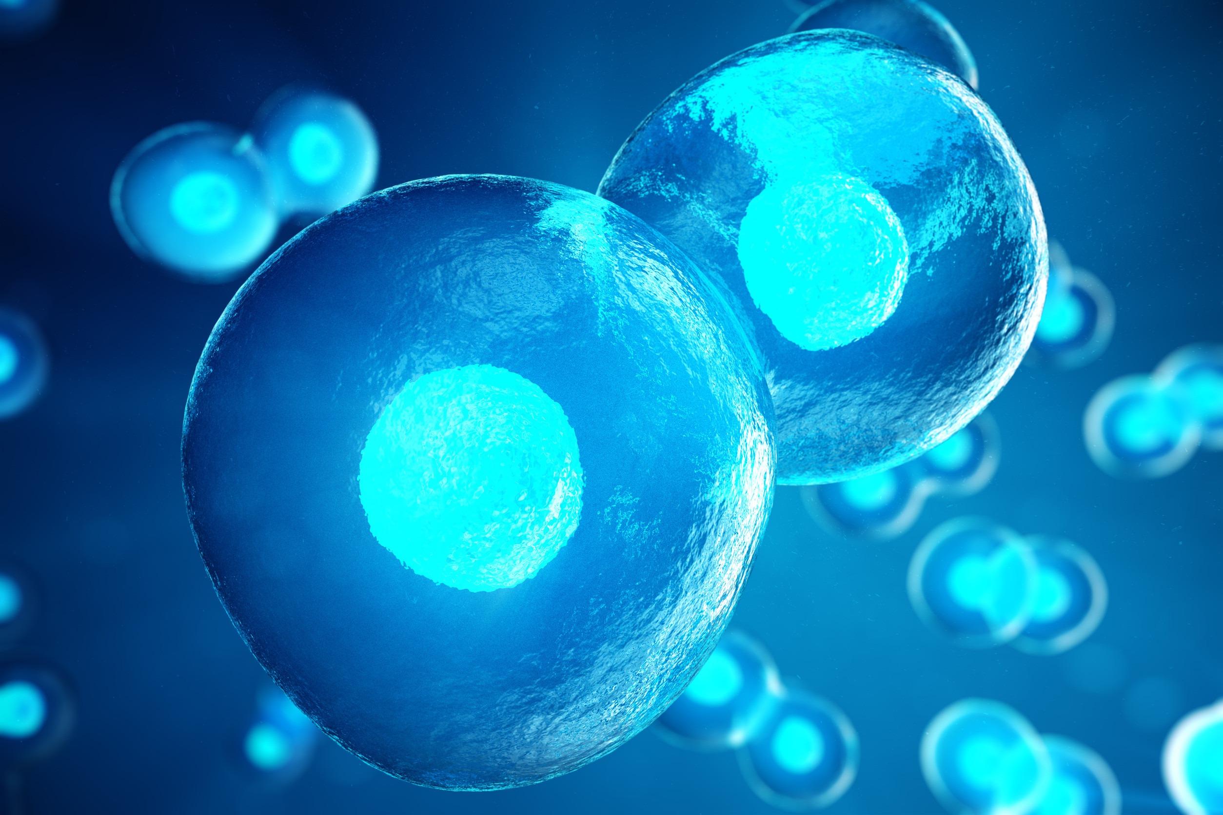
The choice of cells is critical. Cardiomyocytes, the heart’s muscle cells, are essential for contraction and overall function.
A great range of cells are available for CTE. Sources of cardiomyocytes include embryonic stem cells (ESCs), bone marrow-derived mesenchymal stem cells, skeletal myoblasts, endothelial progenitor stem cells, very small embryonic-like stem cells, endogenous cardiac stem cells, induced pluripotent stem cells (iPSCs), and multipotent adult germline stem cells.
The iPSCs, in particular, have garnered attention due to their ability to differentiate into cardiomyocytes while bypassing ethical issues associated with ESCs. Mesenchymal stem cells (MSC) also deserve attention because they can be differentiated into many cell types such as bone, adipose or fat tissue and cartilage. These are found in a great range of tissues and organs. Critically, they are multipotent and extensive proliferation potential. Using adult stem cells allows for autologous cell transplantation.
Scaffolds
Scaffolds provide a three-dimensional structure that supports cell attachment, growth, and organization. These can be made from natural materials like collagen or synthetic polymers such as polylactic acid (PLA). The scaffold’s architecture and material properties are designed to mimic the extracellular matrix (ECM) of cardiac tissue, facilitating cell signaling and integration.
Bioactive Molecules
These include growth factors and cytokines that promote cell survival, proliferation, and differentiation. These molecules are often incorporated into the scaffold or delivered locally to enhance tissue regeneration.
Advances in Cardiac Tissue Engineering
- 3D Bioprinting: One of the most significant advancements is the use of 3D bioprinting to create cardiac tissue. This technology allows for precise placement of cells and materials, creating complex tissue structures that closely mimic the native heart tissue. Bioprinting can produce patient-specific tissues, enhancing compatibility and reducing the risk of rejection.
- Organoids and Microtissues: Researchers have developed cardiac organoids and microtissues that replicate the heart’s structure and function on a smaller scale. These models are invaluable for studying heart development, disease, and drug responses, providing insights that are difficult to obtain from traditional 2D cell cultures or animal models.
- Biomaterials and Smart Scaffolds: Innovations in biomaterials have led to the development of “smart” scaffolds that respond to environmental cues. These materials can change their properties in response to stimuli such as pH, temperature, or mechanical stress, providing dynamic support for tissue growth and function.
- Vascularization: Ensuring adequate blood supply is crucial for the survival and integration of engineered tissues. Techniques such as prevascularization, where blood vessel networks are formed within the scaffold before implantation, and the use of angiogenic factors to promote blood vessel formation post-implantation, have shown promise in addressing this challenge.
Clinical Applications and Challenges
The potential clinical applications of cardiac tissue engineering are vast. These include:
- Myocardial Patches: Engineered myocardial patches can be applied to areas of the heart damaged by myocardial infarction (heart attack). These patches can provide structural support and secrete bioactive molecules to promote repair and regeneration.
- Heart Valves and Conduits: Tissue-engineered heart valves and vascular conduits can replace damaged or diseased structures, improving blood flow and heart function. These constructs are designed to grow and remodel with the patient, potentially reducing the need for multiple surgeries.
- Disease Models and Drug Testing: Engineered cardiac tissues can serve as models for heart disease, allowing for more accurate testing of new drugs and therapies. This can accelerate the development of treatments and reduce reliance on animal models.
Despite these promising applications, several challenges remain:
- Immune Response: Avoiding immune rejection is a significant hurdle. Even with autologous cells (cells derived from the patient), the immune response to scaffolds and other components must be carefully managed.
- Integration and Functionality: Ensuring that the engineered tissue integrates seamlessly with the host tissue and functions correctly is critical. This includes achieving synchronized contraction and proper electrical signaling.
- Scale-Up and Manufacturing: Scaling up the production of engineered tissues to meet clinical demand is complex. Standardizing manufacturing processes to ensure consistency, safety, and efficacy is essential for regulatory approval and widespread clinical use.
Future Directions
The future of cardiac tissue engineering is bright, with ongoing research focusing on several key areas:
- Stem Cell Technology: Advances in stem cell biology, including gene editing techniques like CRISPR, are poised to enhance the efficiency and safety of producing cardiomyocytes.
- Biofabrication: Emerging biofabrication techniques, including advanced bioprinting and nanotechnology, hold promise for creating more sophisticated and functional cardiac tissues.
- Personalized Medicine: Integrating patient-specific data, such as genetic information, with tissue engineering approaches could lead to highly personalized treatments, improving outcomes and reducing complications.
- Regenerative Therapies: Combining tissue engineering with regenerative therapies, such as gene therapy and exosome delivery, could further enhance the heart’s capacity to repair itself.
Cardiac tissue engineering stands at the forefront of regenerative medicine, offering hope for millions affected by heart disease. Through innovative approaches that combine cellular biology, materials science, and bioengineering, this field aims to create functional cardiac tissues that can repair and regenerate the damaged heart. While challenges remain, the ongoing advancements and interdisciplinary collaboration continue to push the boundaries of what is possible, bringing us closer to a future where heart disease can be effectively treated or even cured.
References
Carrier, R. L., Papadaki, M., Rupnick, M., Schoen, F. J., Bursac, N., Langer, R., … & Vunjak‐Novakovic, G. (1999). Cardiac tissue engineering: cell seeding, cultivation parameters, and tissue construct characterization. Biotechnology and Bioengineering, 64(5), pp. 580-589.
Curtis, M. W., & Russell, B. (2009). Cardiac tissue engineering. Journal of Cardiovascular Nursing, 24(2), pp. 87-92.
Eschenhagen, T., Didié, M., Heubach, J., Ravens, U., & Zimmermann, W. H. (2002). Cardiac tissue engineering. Transplant Immunology, 9(2-4), pp. 315-321.
Hirt, M. N., Hansen, A., & Eschenhagen, T. (2014). Cardiac tissue engineering: state of the art. Circulation Research, 114(2), pp. 354-367
Kitsuka T, Takahashi F, Reinhardt J, Watanabe T, Ulziibayar A, Yimit A, Kelly J, Shinoka T. Advances in Cardiac Tissue Engineering. Bioengineering (Basel). (2022) Nov 16;9(11):696. doi: 10.3390/bioengineering9110696. PMID: 36421097; PMCID: PMC9687338.
Liau, B., Zhang, D., & Bursac, N. (2012). Functional cardiac tissue engineering. Regenerative Medicine, 7(2), pp. 187-206.
Nguyen, A. H., Marsh, P., Schmiess-Heine, L., Burke, P. J., Lee, A., Lee, J., & Cao, H. (2019). Cardiac tissue engineering: state-of-the-art methods and outlook. Journal of Biological Engineering, 13, pp. 1-21.
Rabkin, E., & Schoen, F. J. (2002). Cardiovascular tissue engineering. Cardiovascular Pathology, 11(6), pp. 305-317.
Vunjak-Novakovic, G., Tandon, N., Godier, A., Maidhof, R., Marsano, A., Martens, T. P., & Radisic, M. (2010). Challenges in cardiac tissue engineering. Tissue Engineering Part B: Reviews, 16(2), pp. 169-187.
Leave a Reply