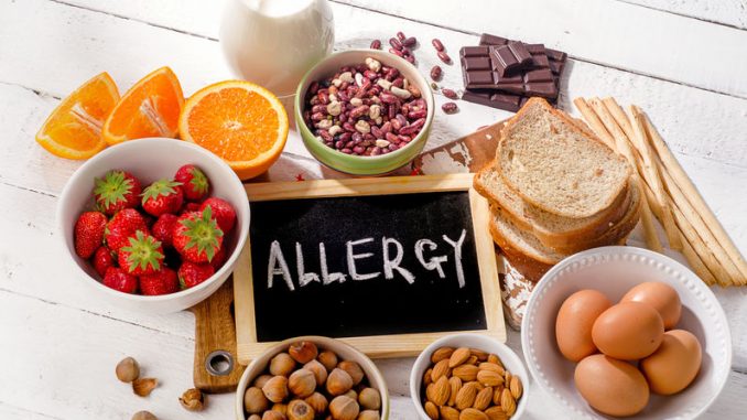
Allergies, especially food allergies have in recent years become a strong focus for research attention because they are so debilitating. Food allergies restrict food use, make life difficult for food manufacturers who must comply with processing and labelling regulations and generally remain a hidden threat to those susceptible to them.
It is a global phenomenon which has seen a steady rise in numbers (Branum and Lukacs, 2009). We know that an accurate estimate of those suffering from allergy generally is hard to predict (Sicherer, 2011) but some authors think the figures is between 2 and 10% of the general population (Chafen et al., 2010). The incidence of allergy correlates relatively well with the increasing popularity of the western diet (Sicherer and Sampson, 2007).
An allergy is essentially an adverse immune response to a foreign substance- the allergen which can be proteins and carbohydrates found in pollen, food ingredients and drugs. A variety of disorders can arise as a result of an allergenic episode which include minor but uncomfortable disorders such as atopic dermatitis, and respiratory and gastrointestinal distress, to the life-threatening anaphylactic responses (Shek and Lee, 2006; Berin and Sampson 2013).
The Mechanisms Behind Food Allergies
When someone comes into repeated contact with any allergen there is a secondary immunological response. This is often in complete disproportion to exposure and leads to the very serious health conditions that were described earlier.
Gell and Coombs developed a classification system for the four basic types of allergy. These are Type I through to Type IV. Each one forms through different immunological mechanisms.
Allergy Type I
The most frequently diagnosed type of allergy.
It involves an immediate, immunoglobulin E (IgE)- mediated hypersensitivity. This occurs by attachment of a specific IgE immunoglobulin to mastocyte (mast) cells. This type of immunoglobulin is increasingly released after repeated exposure to allergens. The binding of IgE leads to gradual binding of receptors and induces cross-linking of the IgE-antibody by the specific allergen.
Mast cell degranulation follows with the release of various mediators of the immune response. We see a sudden surge in prostaglandins, leukotrienes, thromboxanes and others. The mediators cause vasodilation, contraction of smooth muscle, a rise in mucus secretions, nerve endings are irritated.
Six to eight hours after contact with the allergen, eosinophils, neutrophils and lymphocytes migrate to the spot of initial allergen entry.
Allergen Type II
Gell and Coombs describe this as ‘antibody-mediated cytotoxicity’. This is triggered by damage to the hapten-binding cells, These cells become antigens and induce production of another class of immunoglobulins called the IgM and IgG antibodies. These attach to haptens on the cell membrane and activate complement proteins which leads to cell lysis
Allergen Type III
This is when an acute inflammatory response occurs. Large numbers of antibody-antigen complexes accumulate in tissues. There is a extreme damage to cells and tissue. It is a phenomenon associated with ulcerative colitis.
Allergen Type IV
The delayed hypersensitivity reaction when an antigen interacts with an antigen-specific lymphocyte. These release inflammatory and toxic substances that attract white blood cells to swarm.
Biochemical Mechanisms For Food Allergies
Clearly, there is a need to understand the biochemical mechanisms that underly the condition.
The mechanisms are slowly being unravelled but they are not completely understood (Berin and Sampson, 2013). A food allergy is mediated by the activity of certain cells involved in immunology, the T-helper 2 cells (Th2 cells) which is a T-cell subtype. The Th2 cells, when activated. stimulate the B cells which generate the allergen-specific immunoglobulin E (IgE) (Georas et al., 2005).
Secondary oral exposure to a specific food antigen subsequently causes a wide range of symptoms, including systemic and localized anaphylaxis (Berin and Sampson 2013). During the early responses of food allergy, allergens are taken up by antigen-presenting cells, which present selected peptides on the major histocompatibility complex (MHC) class II molecules to activate naïve helper T cells (Th0 cells) into Th cells. This then results in the activation of Th2 cells through the transcription factor GATA-binding protein 3, which then produces cytokines such as IL-4, IL-5, and IL-13 (Hammad and Lambrecht, 2006). The cytokines incidentally are small proteins of about 5 to 20 kDa that are needed in cell signaling. They modulate the balance between humoral and cell-based immune responses.
In contrast, Th1 cells primarily produce IFN-γ and IL-12 via the transcription factor T-bet (Glimcher, 2007). Th1 cells release mediators that contribute to the suppression of IgE production by inhibiting Th2 cell proliferation (Murosaki et al., 1999). Therefore, maintaining a balance between Th1 cells and Th2 cells is critical for preventing and treating food allergy.
References
Berin, M.C., Sampson, H.A. (2013) Mucosal immunology of food allergy. Curr. Biol. 23(9): R389–R400 (Article)
Branum, A.M., Lukacs, S.L. (2009) Food allergy among children in the United States. Pediatrics 124(6) pp. 1549–55
Chafen, J.J., Newberry, S.J., Riedl, M.A., Bravata, D.M., Maglione, M., Suttorp, M.J., Sundaram, V., Paige, N.M., Towfigh, A., Hulley, B.J., Shekelle, P.G. (2010) Diagnosing and managing common food allergies: a systematic review. J.A.M.A. 303(18) pp. 1848–56
Georas, S.N., Guo, J., De Fanis, U., Casolaro, V. (2005) T-helper cell type-2 regulation in allergic disease. Eur. Respir. J. 26(6) pp. 1119–37
Glimcher, L.H. (2007) Trawling for treasure: tales of T-bet. Nat. Immunol. 8(5) pp. 448–50.
Hammad, H., Lambrecht, B.N. (2006) Recent progress in the biology of airway dendritic cells and implications for understanding the regulation of asthmatic inflammation. J. Allergy Clin. Immunol. 118(2) pp. 331–6.
Murosaki, S., Muroyama, K., Yamamoto, Y., Kusaka, H., Liu, T., Yoshikai, Y. (1999) Immunopotentiating activity of nigerooligosaccharides for the T helper 1-like immune response in mice. Biosci. Biotechnol. Biochem. 63(2) pp. 373–8.
Shek, L.P., Lee, B.W. (2006) Food allergy in Asia. Curr. Opin. Allergy Clin. Immunol. 6(3) pp. 197–201
Sicherer, S.H. (2011) Epidemiology of food allergy. J. Allergy Clin. Immunol. 127(3) pp. 594–602
Sicherer, S.H., Sampson, H.A. (2007) Peanut allergy: emerging concepts and approaches for an apparent epidemic. J. Allergy Clin. Immunol. 120(3) pp. 491–503; quiz 4–5


Leave a Reply